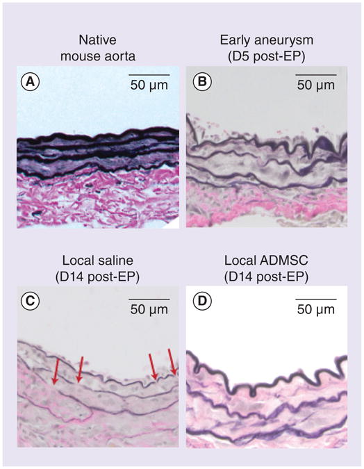
Figure 4. Qualitative examination of elastin.

Images from native aorta, early aneurysm, untreated aneurysm and local ADMSC treatment groups are shown after Verhoeff–Van Gieson staining (n = 2 all groups).
ADMSC: Adipose-derived mesenchymal stem cell; D: Day; EP: Elastase perfusion.
