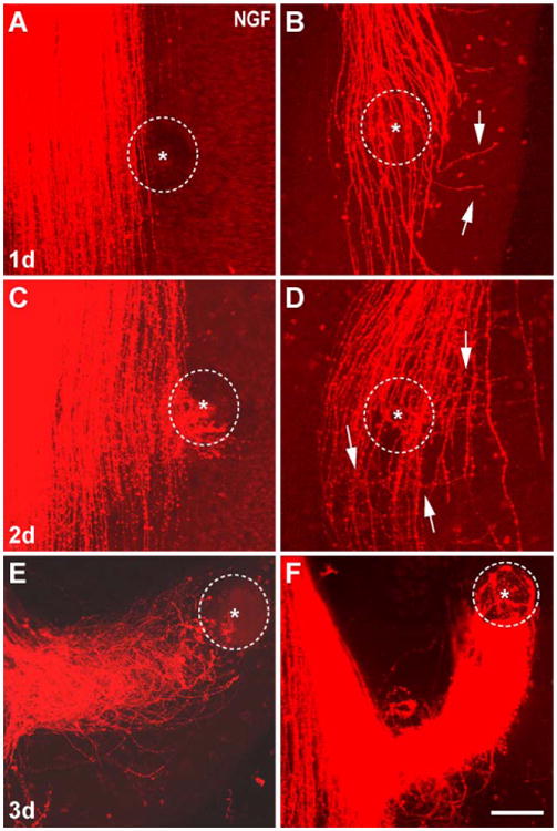
Fig. 3.

The effects of NGF bead over a three-day culture period. (A) After 24 h most cases did not show any axonal response to the NGF bead. (B) When NGF bead was placed over the trigeminal tract on the ventricular side of the explant, a distinct defasciculation and turning response of axons that have reached this level is seen. Some axons tipped with growth cones (arrows) leave the tract and grow away from the bead. After 48 h in culture, many axons show attraction response to the NGF bead (C). (D) A case with an NGF bead over the tract. A clear turning of the leading edge of immature axons is evident (arrows). After 3 days in culture, central trigeminal axons extended all the way to the most laterally placed beads, forming spiraling funnels (E, F). In these two exemplary cases the central trigeminal tract is located to the left of the micrographs. Dashed circles outline the beads and asterisks mark the center of the beads. Scale bar=150 μm.
