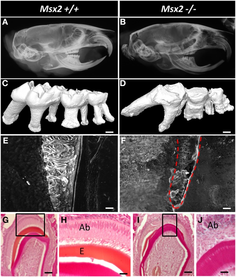
Figure 3.
Dental phenotype of Msx2−/− mice with reference to wild-type animals (Msx2+/+). (A,B) Microradiographs of the whole heads of 3-month old mice showing craniofacial and teeth dysmorphology; indeed craniofacial morphogenesis is under the control of MSX2 (Simon et al., 2014). Msx2−/− mutant mice present a non-isometric overall craniofacial size decrease; the teeth exhibit crown and root dysmorphology with altered enamel, enlargement of the pulp cavity, short and curved roots with abnormal orientations, and reduced curvature of the incisor. The third molar showed impaired eruption and the most severe phenotype. (C,D) 3D reconstruction of mouse molars revealed the absence of cuspid relief and severe generalized enamel hypoplasia with irregular surface. Msx2−/− mice displayed complex radicular morphology (Aïoub et al., 2007). (E,F) Scanning electron microscopy of the first molar mandible illustrates a severe reduction in enamel thickness. Enamel in Msx2−/− animals shows the absence of enamel prisms, replaced by an amorphous layer (Molla et al., 2010); scale bars: 10 μm. (G–J) Histological analysis of mouse molar enamel reveals hypoplastic amelogenesis imperfecta in Msx2−/− mice. This feature is related, after a correct ameloblast differentiation process, to a secondary inability of ameloblasts to secrete the enamel matrix which would mineralize. Ameloblast cells in these animals lose their polarization, become rounded and isolated, and finally disappear (Ab, ameloblast; E, enamel —scale bars: G, I: 100 μm; H, J: 40 μm).

