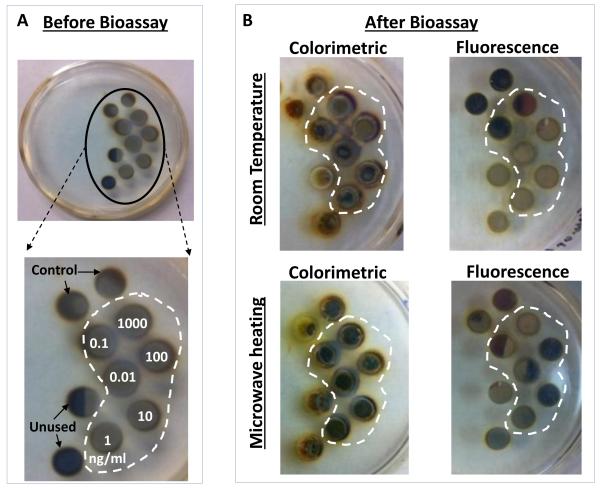
Fig. 4.
Real-color photographs of circular bioassay platforms (a) before bioassays: the numbers indicate the wells assigned to the specific concentration of p53 used. Control: control bioassay without p53. Unused: water only (no bioassay), and (b) after the completion of colorimetric and fluorescence based bioassays at room temperature and using MAB. Note that the silicon isolators were used in all bioassays but not shown here for clarity of the pictures.

