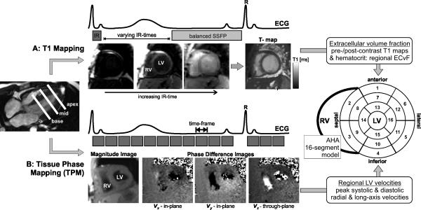
Figure 1.
A: Combined T1 and Tissue Phase Mapping (TPM) for the comprehensive segmental evaluation of myocardial extracellular volume fraction (ECV) and function (3-directional left ventricular (LV) velocities). A: T1 mapping is based on diastolic balanced SSFP imaging with different inversion recovery (IR) times. B: Tissue phase mapping is based on ECG gated black-blood prepared phase contrast MRI with three-directional velocity encoding. Both T1 mapping and MVM were acquired in short axis orientation (base, mid, apex) during breath-holding. Results from both measurements were mapped on the AHA 16-segment model for direct segmental comparison of ECV and LV peak velocities.

