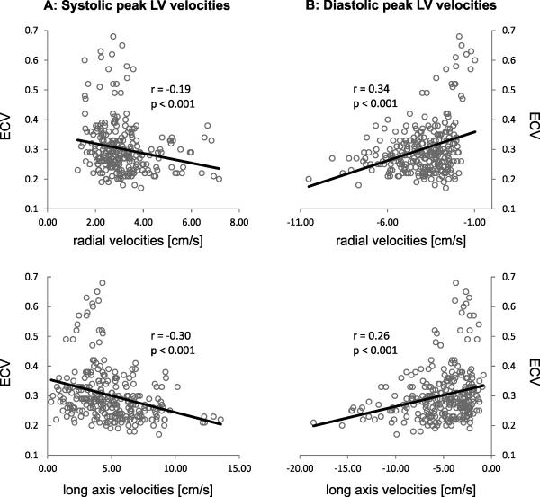
Figure 3.
Correlation analysis between segmental systolic (A) and diastolic (B) LV peak velocities and regional myocardial extracellular volume fraction (ECV) in patients with nonischemic heart disease and preserved LVEF. The individual graphs show the results of the correlation of regional ECV with myocardial velocities for 19 patients with preserved LVEF on a segment-by-segment basis. For each subject, data form all 16 segments were included, resulting in a total of 19 x 16 = 304 data points (ECV - myocardial velocity pairs) for each graph.

