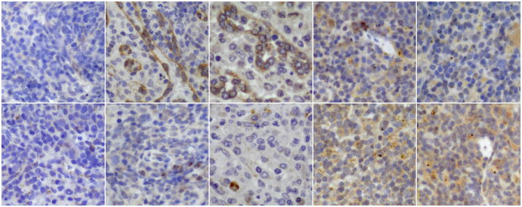
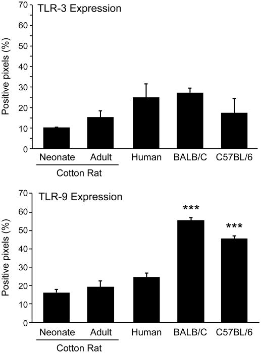
Figure 1. TLR-3 and TLR-9 expression in cotton rat, human and mouse spleen cells.
Paraffin-embedded sections of spleens (three per group) from neonatal cotton rats (3 days old), adult cotton rats (7 weeks), adult humans (20 to 40 years old), BALB/C and C57BL/6 mice (6 weeks old) were stained with cross-reactive polyclonal sera specific for human TLR-3 and TLR-9.
A. TLR-3 specific stain (brown) is shown in the top panel, and TLR-9 specfic stain (brown) in the bottom panel (400×). From left to right neonatal and adult cotton rat spleen, human spleen, BALB/C and C57BL/6 mouse spleen.
B. Positive pixels were quantified with the Aperio image scope positive pixel counter algorism. The number of asterisks denotes the level of statistical significance (***p < 0.001).


