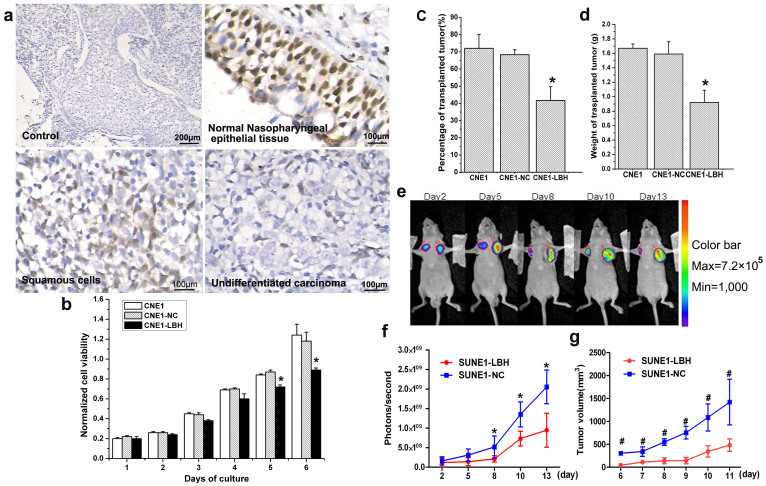
Figure 1. LBH is expressed in NPC tissues and inhibits the proliferation and growth of xenografted NPC tumors in BALB/c nude mice.
(a) Negative control (no primary antibody) of nasopharyngeal epidermal tissue showed no positive staining in IHC. Almost all normal epidermal cells in normal nasopharyngeal epidermal tissue exhibited strong positive LBH staining. Partially cancerous cells of the squamous cell NPC tissue type exhibited moderately positive LBH staining. Almost all cancer cells of the nasopharyngeal undifferentiated NPC tissue type were negative for LBH staining. (b) MTT assay showed that LBH expression inhibited CNE1 cell proliferation (*P < 0.05 vs. CNE1 or CNE1-NC group). (c) Tumor-formation rate after CNE1, CNE1-NC, or CNE1-LBH cell (5 × 10⁶) injection into the axilla of BALB/c nude mice was 72%, 68% and 42%, respectively (*P < 0.05 vs. CNE1 or CNE1-NC group). (d) The average weight of transplanted tumor in nude mice injected with CNE1, CNE1-NC, and CNE1-LBH cells was 1.67 g, 1.59 g, and 0.92 g, respectively. (*P < 0.05 vs. CNE1 or CNE1-NC group) (e) Representative image of the inhibition effect on SUNE1 tumor xenograft growth by LBH expression detected by bioluminescent imaging. SUNE1-NC and SUNE1-LBH cells were injected subcutaneously into the left and right armpits, respectively, in nude mice. (f) Photons/sec analysis showed that the growth of the SUNE1-LBH xenografted tumors was slower than SUNE1-NC control tumors (*P < 0.05). (g) The volume of transplanted tumors in nude mice also showed that the SUNE1-LBH grew slower than the control SUNE1-NC tumors (#P < 0.01).

