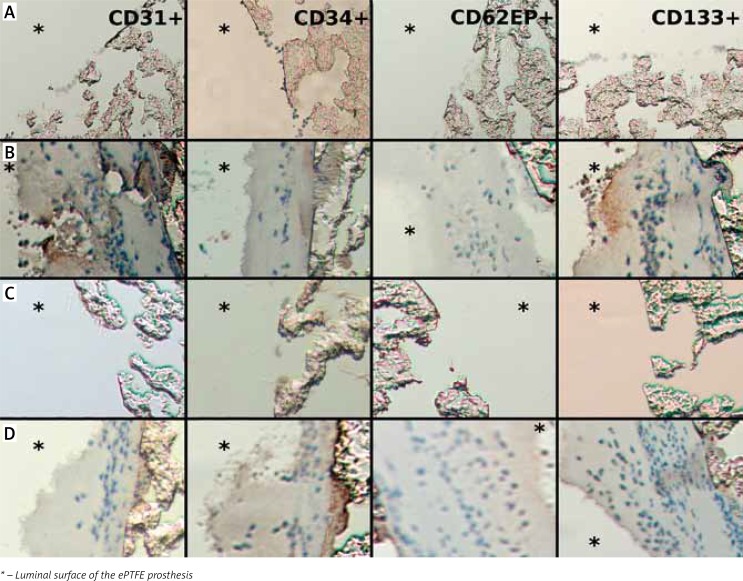
Fig. 3.
Results of immunostaining with anti-CD31, anti-CD34, anti-CD62EP, and anti-CD133 antibodies (magnification 200×). All grafts after 120 min of perfusion. No positive staining for EPC visible in any grafts or any antibody type. A) Dry control ePTFE vascular graft. B) Dry anti-CD34-coated ePTFE vascular graft. C) Wet control ePTFE vascular graft. D) Wet anti-CD34-coated ePTFE vascular graft

