
Figure 4.
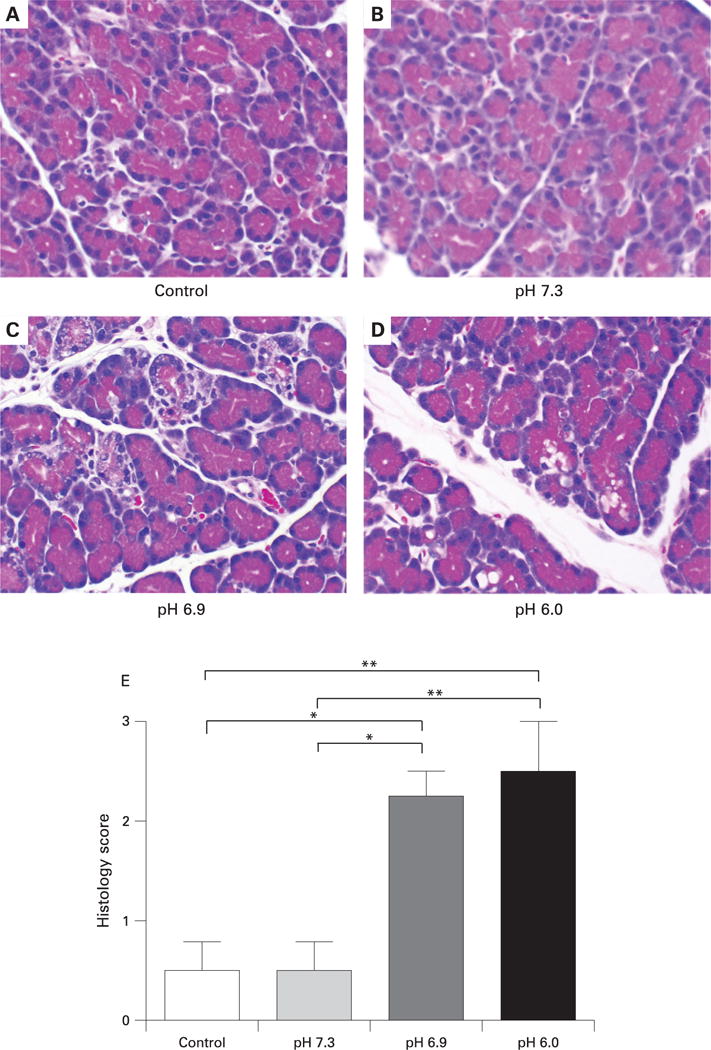
Effect of high pressure intraductal injection of contrast solutions on pancreatic histology.
(A–D) Representative photomicrographs of pancreatic tissue collected 24 h after injection of solutions of different pH into the pancreatic duct. Solutions at pH 6.9 produced histological disruption and oedema (C). These changes were more marked and accompanied by neutrophil infiltration in tissues from animals injected with solution at pH 6.0 (D). Magnification: ×6250. (E) Histological scoring of pancreatic sections was performed by an observer blinded to the study design. *p<0.05. **p<0.01.