
Figure 7.
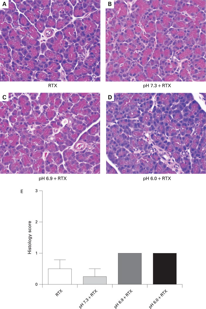
Effects of resiniferatoxin (RTX) on pancreatic histology when co-injected into the pancreatic duct with contrast solutions of different pH.
(A–D) Photomicrographs of representative pancreatic tissue collected 24 h after injection of solutions of different pH into the pancreatic duct. Magnification: ×6250. (E) Histological scoring of pancreatic sections was performed by an observer blinded to the study design. *p<0.05, **p<0.01.