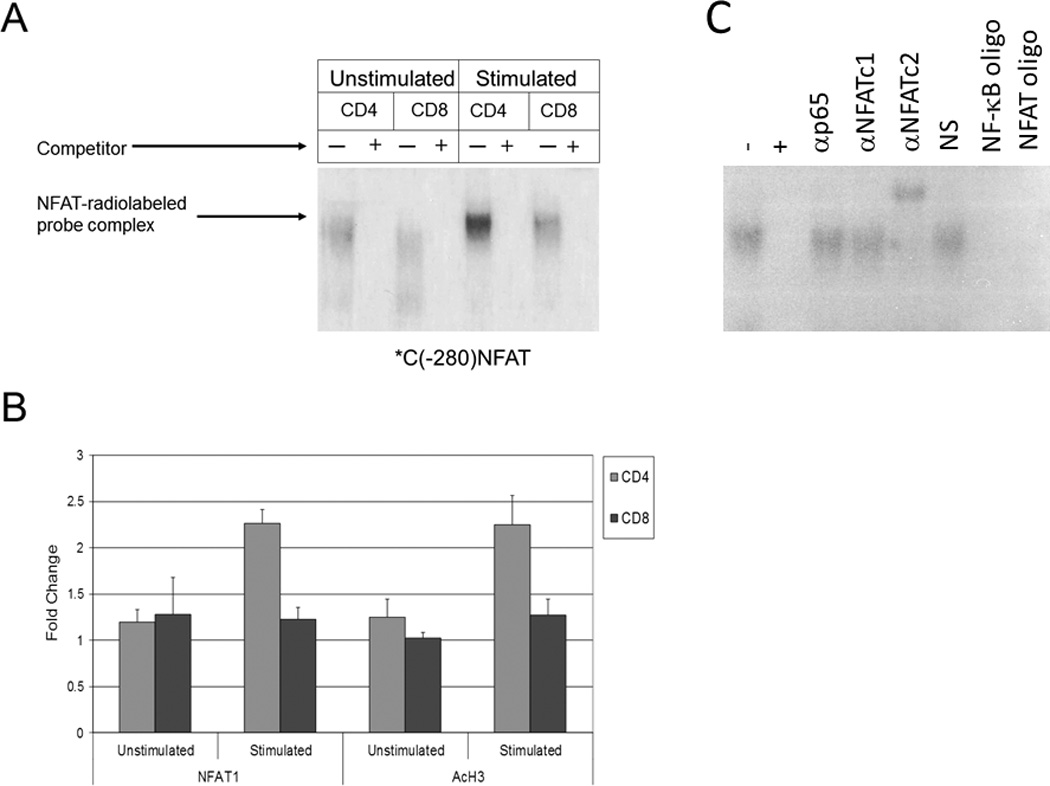
Figure 3. Increased NFAT activity is found in CD4+ vs. CD8+ T cells at the CTLA-4 promoter.

a) Protein extracts from unstimulated and stimulated CD4+ and CD8+ T cells were incubated with a radiolabelled probe representing an NFAT binding site in the CTLA-4 promoter (*C(−280)NFAT). Subsequently, mixtures were then incubated with or without excess competitor unlabeled probe (+ or −) and NFAT1 binding was assayed via 4% polyacrylamide gel electrophoresis. b) CHIP assays were performed on the CTLA-4 promoter region using anti-NFAT1 (left panel) and anti-acetylated histone H3 (AcH3, right panel) antibodies in unstimulated and stimulated CD4+ and CD8+ T cells. Normalization for equal cell numbers was 10% of the input DNA. Results in each panel are averages from two independent experiments. c) Gel shift mobility assay (EMSA) was performed as described above with our without excess competitor unlabeled probe (+ or −) and gel shift was assessed using antibodies to the p65 subunit of NF-κB, NFAT1, and NFAT2. Normal serum (NS), NF-κb oligonucleotides (NF-κb oligo) and NFAT oligonucleotides (NFAT oligo) were used as additional controls.
