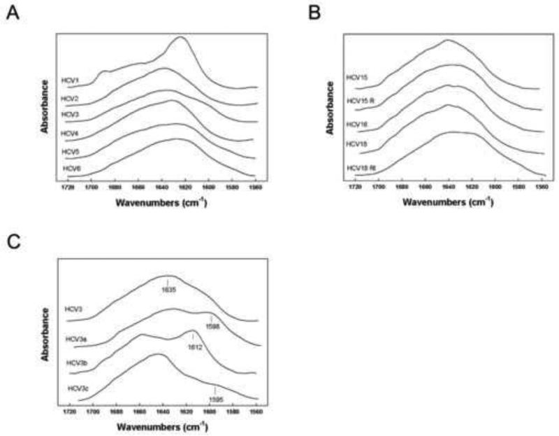
Figure 3.
FTIR spectroscopy analyses of HCV peptides. A and B. FTIR spectral analysis of unlabeled HCV peptide secondary structure in deuterated 10 mM phosphate buffer pD 5.6. C. FTIR spectral analysis of HCV3 13C isotope enhanced peptide secondary structure in deuterated 10 mM phosphate buffer pD 5.6. HCV3 is unlabeled peptide; HCV3a is isotopically enhanced at residues Phe3 and Val5; HCV3b is isotopically enhanced at Gly6 and Leu7; and HCV3c is isotopically enhanced at Tyr10.

