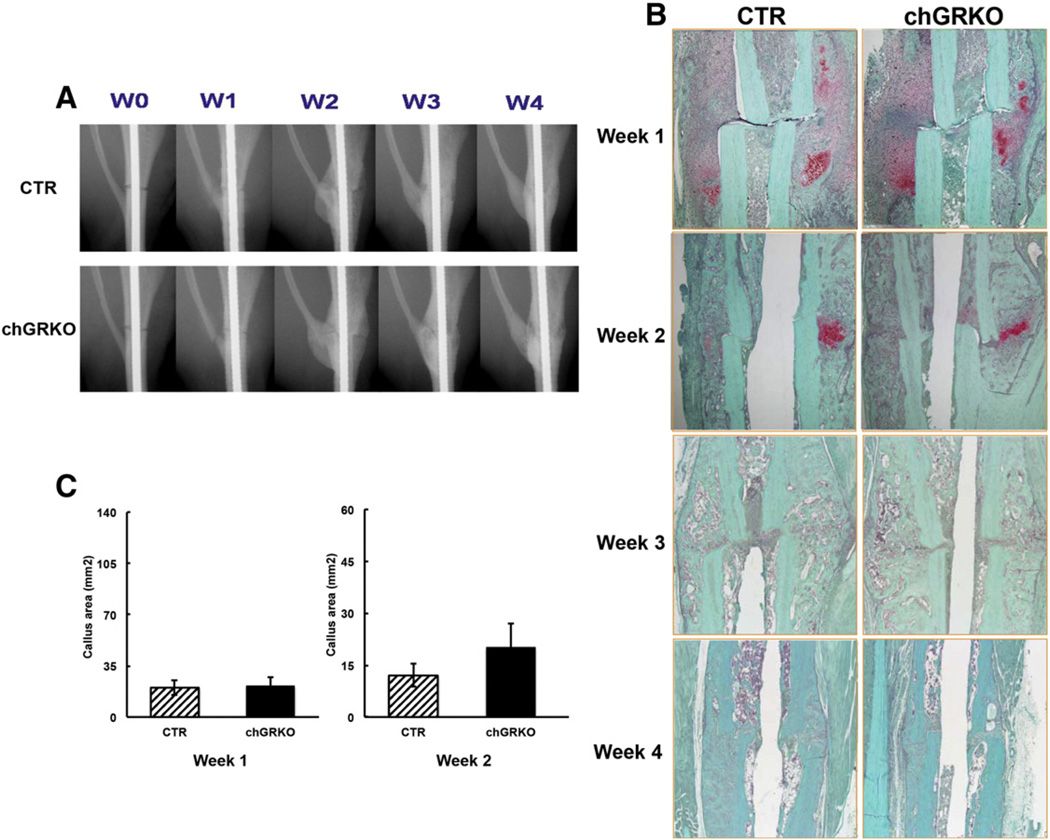
Fig. 6.
X-ray and histological evaluations of the images of diaphyseal fracture healing. A, Representative X-ray images of tibial fractures over the course of the diaphyseal fracture study; B, Representative histological images of tibial fractures displayed less displacement of fractured bone at all four time points than corresponding metaphyseal fractures. In addition, chGRKO mice demonstrated similar fracture repair compared to the CTR mice; C, histomorphometric analysis at weeks 1 and 2 revealed similar cartilaginous callus area in chGRKO and CTR mice.

