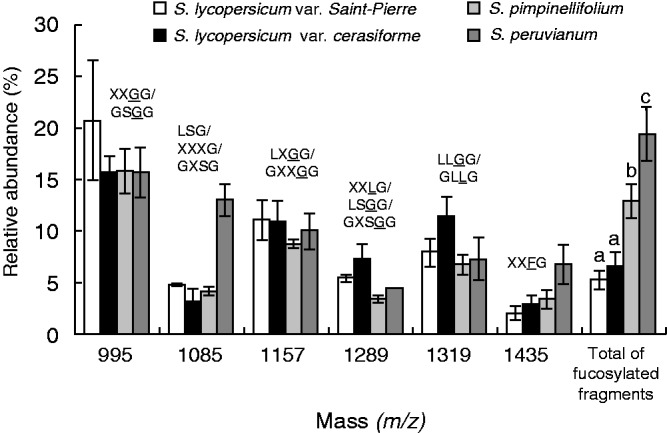
Fig. 5.

Comparison of the oligosaccharide profile of the main endo-glucanase-sensitive XyG fragments between S. lycopersicum var. Saint-Pierre, S. lycopersicum var. cerasiforme ‘wva106’, S. pimpinellifolium and S. peruvianum pollen tubes. The possible structures of XyG fragments are shown according to the nomenclature proposed by Fry et al. (1993) and shown in Fig. 1. Underlined structures represent O-acetylated side chains. The mean relative abundance is the average of three independent biological samples (n = 3 ± s.d.). Different letters indicate significant differences (P < 0·05).
