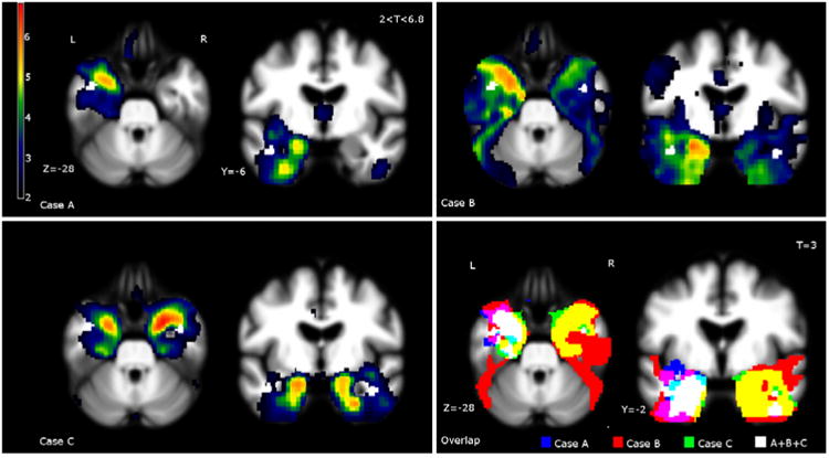
Figure 2.

Single-subject VBM and overlap of the three cases.
Case A: Bilateral, left greater than right, anterior temporal atrophy extending to the hippocampus and orbitofrontal areas. Case B: Bilateral, left greater than right, anterior temporal lobe atrophy. Case C: Bilateral, right greater than left, atrophy in the anterior temporal lobes and amygdala. Moderate bilateral a trophy in the orbitofrontal cortex, anterior insula, and parahippocampus. Overlap: To demonstrate overlap of damage in language dominant regions, Case B has been L-R flipped because of his right-dominant language organization. All three patients had relative sparing in the dominant (shown as left) lateral superior temporal lobe, including the superior and middle gyri, and uniform atrophy in the medial temporal cortex, including the amygdala, insula, and parahippocampalgyri.
