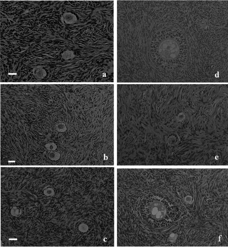
Fig. 2.

Histological analysis of fresh, nonvitrified and vitrified-warmed pre-antral follicles. (a) Primordial and primary follicles in fresh human ovarian cortical tissue. (b) Primordial and primary follicles in nonvitrified human ovarian cortical tissue after 6 h of transportation. (c) Primordial and primary follicles in nonvitrified human ovarian cortical tissue after 18 h of transportation. (d) Secondary follicles in nonvitrified human ovarian cortical tissue after 18 h of transportation. (e) Primary follicles in vitrified-warmed human ovarian cortical tissue after 18 h of transportation. (f) Primordial to secondary follicles in vitrified-warmed human ovarian cortical tissue after 18 h of transportation. (× 200; H-E staining). The scale bars represent 20 μm.
