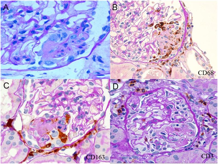
Figure 1.
Neutrophils, CD68+ and CD163+ macrophages, and rare CD3+ T cells are localized at sites of fibrinoid necrosis. (A) Segmental fibrinoid necrosis (FN) with capillary basement membrane perforation, fibrin thrombosis and exudation, polymorphonuclear neutrophils, mononuclear cells, and swollen extracapillary cells. (B) Segmental FN showing localization of CD68+ cells around and within the lesion. Pericapsular CD68+ cells are also evident. (C) Segmental FN showing localization of CD163+ cells in the lesion. Pericapsular CD163+ cells are prominent. (D) CD3+ cells were located primarily in the pericapsular region in glomeruli with segmental FN.

