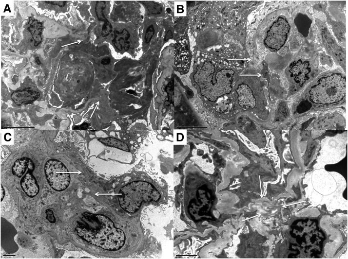
Figure 6.
Ultrastructural features of minute exudative fibrinoid necrosis, glomerular basement membrane perforations, and attenuations. (A) Exudative fibrinoid necrosis with glomerular basement membrane (GBM) perforation (arrows), fibrin thrombus attached to endothelium, and fibrin exudate and mononuclear inflammatory cells with long processes attached to fibrin sheaves and thrombus. (B) Early lesion with a minute GBM perforation (arrows), intracapillary mononuclear cells, and swollen epithelioid cells in Bowman’s space. (C) Endocapillary mononuclear cells extending processes through a minute GBM perforation (arrows). (D) Wrinkled redundant GBM at the mesangium (arrows) with cellular processes extending from the capillary lumen (lower middle) and mesangial cells with numerous processes extending toward the redundant GBM.

