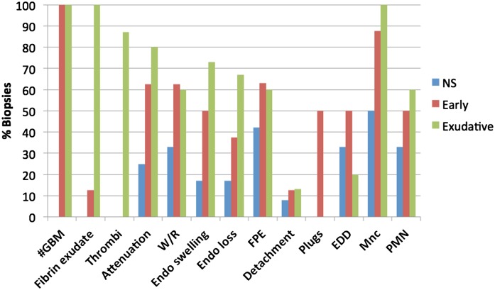
Figure 7.
The distribution of ultrastructural lesions seen in biopsies with pauci-immune necrotizing GN (PNGN). Exudative lesions of fibrinoid necrosis (n=15) are shown in green, early lesions (n=8) are shown in red, and glomeruli without apparent diagnostic abnormalities (nonspecific [NS], n=12) are shown in blue. Endothelial injury, glomerular basement membrane attenuation and redundancy, podocyte injury, and mononuclear and polymorphonuclear neutrophil infiltrates are common accompaniments of severe glomerular injury and also present in less injured glomeruli from biopsies with PNGN. Lesions designated NS may, in fact, be the earliest recognizable lesions of PNGN in the correct clinical and pathologic context. EDD, electron dense deposit; FPE, podocyte foot process effacement; #GBM, glomerular basement membrane perforation; Mnc, mononuclear cell; PMN, polymorphonuclear neutrophil; W/R, wrinkling/redundancy.

