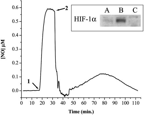
Fig. 8.
Electrochemical detection of NO in the presence of MCF7 cells. MCF7 cells were added in suspension (3 × 10⁶/ml) as described in Materials and Methods. O2 was monitored continuously for 120 min (data not shown). Cells were isolated, and proteins were immunoblotted for HIF-1α. (A) Normoxia (≈21% O2); (B) hypoxia (<1% O2); (C) intermediate (≈10% O2). Representative NO electrode data for condition C are shown (n = 3). Sper/NO (100 μM) was added to the chamber (1). MCF7 cells were added after a steady-state NO level was achieved (2).

