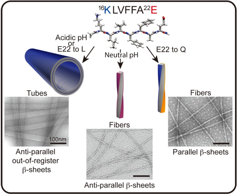
Figure 2.
Aβ [16,17,18,19,20,21,22] peptide assembles into distinct morphologies depending on sequence and conditions. In the cartoons above, blue represents the positively charged lysine (K), red the negatively charged glutamate (E), and orange the uncharged glutamine (Q). Two of the four faces of the fiber in the anti-parallel β-sheets present lysine and glutamate residues on the surface (purple), whereas the fibers with parallel β-sheets have a lysine surface and a glutamine surface. (Unpublished EM images from members of Lynn Lab.)

