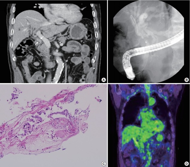
Fig. 1.

Study findings before surgery. (A) A portal phase image of a dynamic computed tomography scan shows luminal narrowing of the mid common bile duct (CBD) with diffuse wall thickening and enhancement (white arrows). (B) A filling defect in the CBD with proximal dilatation is noted upon endoscopic retrograde cholangiopancreatography (ERCP). (C) ERCP biopsy of the tumor reveals a few atypical cells with ulcer detritus, suggestive of adenocarcinoma. (D) Positron emission tomography shows no significant abnormal uptake.
