
Fig. 2.
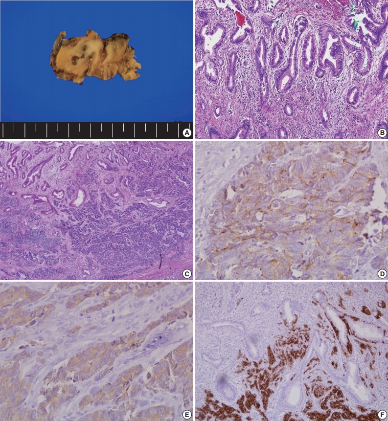
Pathologic findings after surgery. (A) Gross cross-sectional view of the tumor after fixation. (B) On the surface of the tumor, the tumor cells are composed of moderately differentiated adenocarcinoma. (C) The infiltrative tumor cells are small and round with hyperchromatic nuclei and scant cytoplasm. Immunohistochemical analysis for chromogranin (D), synaptophysin (E), and CD56 (F).