
Figure 3. Representative LeBail fit of an X-ray diffraction spectrum of siderite II at 90 GPa and room temperature.
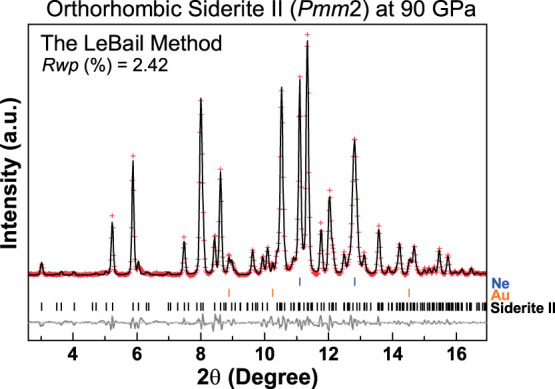
The sample was temperature-quenched to room temperature from 2200 K at 90 GPa. Pluses: measured powder diffraction pattern after background subtraction; black solid curve: refined profile; grey solid curve: residual between the observation and the refinement; vertical ticks: Ne (blue), Au (orange), and siderite II (black).