
Figure 2. Downregulation of miR-125a, miR-574-3p, and miR-877 in 4-hydroxytamoxifen (OHT)-resistant MCF-7 cells (OTHR cells) and clinical breast cancer tissues, and knockdown of miR-574-3p–promoted MCF-7 cell growth in the presence of OHT.
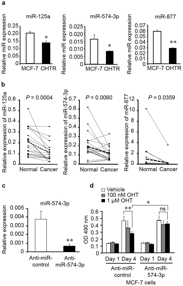
(a) Expression levels of miR-125a, miR-574-3p, and miR-877 in MCF-7 cells and OHTR cells were determined by quantitative PCR (qPCR) and normalized to RNU48 levels. Data are presented as mean ± SE. *P < 0.05; **P < 0.01. (b) Decreased expression levels of miR-125a, miR-574-3p, and miR-877 in breast cancer tissues compared with those in paired adjacent normal tissues. (c) Knockdown efficiency of anti-miR-574-3p. MCF-7 cells were transfected with anti-miR-574-3p or negative control for 48 h. miR-574-3p levels were determined by qPCR and normalized to RNU48 levels. Data are presented as mean ± SE in triplicates. **P < 0.01. (d) Knockdown of miR-574-3p significantly increased MCF-7 cell growth in the presence of OHT. Cells were transfected with anti-miR-574-3p or negative control for 12 h, and then cell viability was analyzed using the MTS cell proliferation assay at 1 and 4 days after transfection. Data are presented as mean ± SE, in triplicate; *P < 0.05; **P < 0.01; ns, not significant.