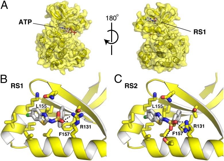
Fig. 2.
Structures of the RS compounds bound to the PIF pocket of PDK1. (A) Structure of the PDK1-RS1 complex. PDK1 is shown as a yellow surface, and both ATP and RS1 are shown as white sticks colored by heteroatom. The relative orientation of the ATP-binding site and the PIF pocket is depicted. (B) Close-up view of the PDK1–RS1 interaction. (C) Close-up view of the PDK1–RS2 interaction.

