Fig. 3.
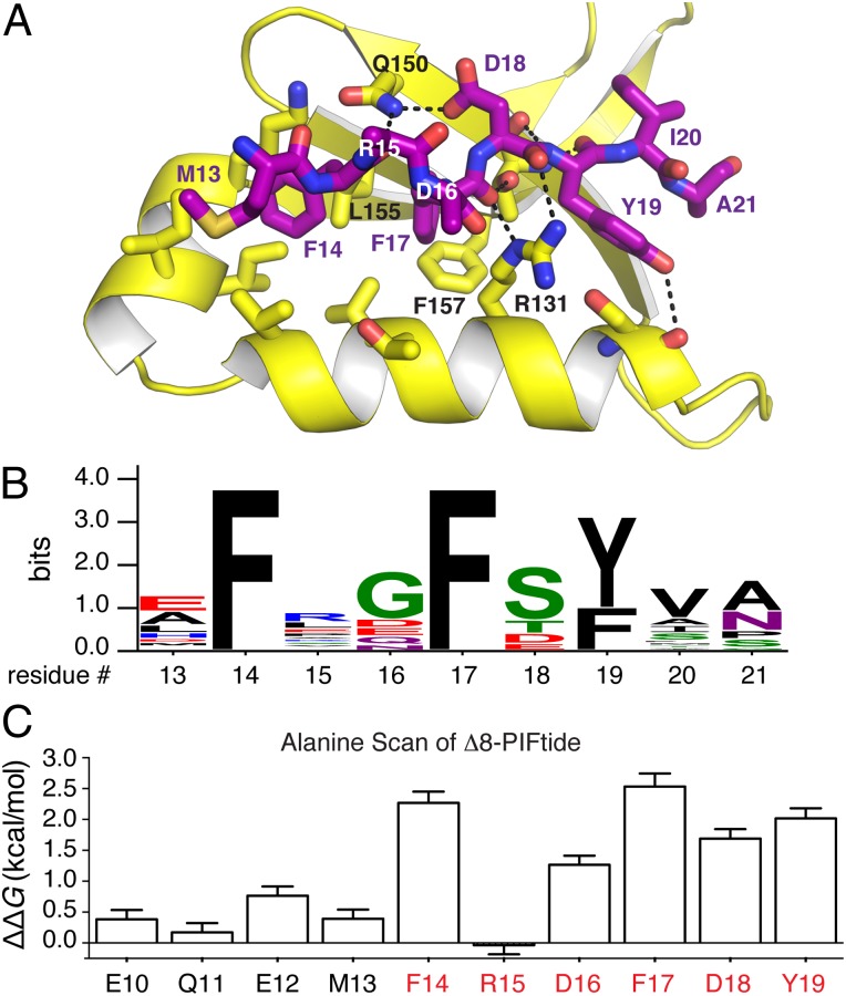
Structural and energetic analysis of the PDK1–PIFtide interaction. (A) Structure of the PDK1-PIFtide complex. PDK1 is in yellow, and PIFtide is shown as purple sticks. (B) Sequence logo depicting the consensus HM from 28 AGC kinases known or inferred to interact with PDK1. The consensus sequence is xFxxF[−](Y/F)(V/A/I)x, where [−] indicates a negatively charged residue (Asp, Glu, or phosphorylated Ser/Thr). (C) Hot spot analysis of the PDK1–PIFtide interaction. Residues 10–19 of PIFtide9–23 were subjected to alanine scanning. The red-colored residues red constitute the HM. Energetic contributions were determined from the Ki values for mutant peptides using the formula ΔΔG = 1.4(kcal/mol)*log(Ki,mut/Ki,wt).

