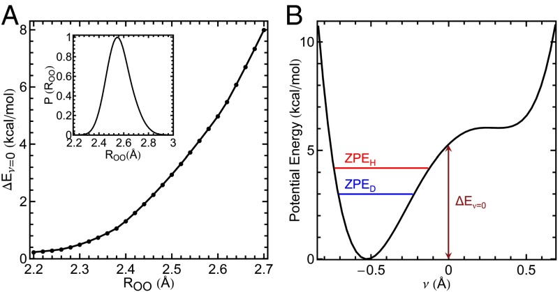
Fig. 4.
Comparison of the energy required to share a proton between residues as a function of the hydrogen-bond donor–acceptor O–O distance compared with the ZPE. (A) as a function of the O–O distance between O16 and O57 using the tyrosine triad geometry from a crystal structure (details in SI Materials and Methods, section D). A, Inset shows the probability distribution of obtained from the AI-PIMD simulation of with ionized Tyr57. The probability is normalized by the maximum value. (B) Potential energy as a function of the proton transfer coordinate ν for = 2.6 Å, indicating values for H and D (O–H and O–D) ZPEs ( and , respectively). The position of Tyr32 is fixed as the proton H16 is scanned along .

