Fig. 1.
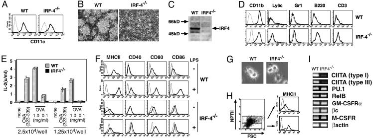
Impaired DC development in the IRF-4–/– BM culture with GM-CSF. (A) The CD11c expression on nonadherent cells from GM-CSF BM cultures at day 10 was analyzed by flow cytometry. (B) Photographs of the cultures by phase contrast microscopy, taken at day 10. (C) The expression of IRF-4 in the CD11c+ cells was assessed by immunoblotting. Lysates from 2.5 × 10⁵ cells were subjected to electrophoresis. (D) The lineage marker expression on the CD11c+ cells was analyzed by flow cytometry. (E) The antigen-presenting ability of the CD11c+ cells for whole OVA and its peptide (323–339 amino acid residues) to OVA-specific CD4+ T cells was examined. (F) MHC-II and costimulatory factor expression by the nonstimulated and LPS-stimulated CD11c+ cells was examined. (G) The morphology of LPS-stimulated CD11c+ cells was observed by phase contrast microscopy. (H) Wild-type and IRF-4–/– BM cells were cocultured. After 10 days, the expression of MHC-II on the cells was analyzed. NPTII expression by the wild-type and the IRF-4–/– cells was distinguished by flow cytometry with anti-MHC-II and anti-NPTII. (I) The expression of several transcription factor and cytokine receptor genes involved in DC development was analyzed by RT-PCR.

