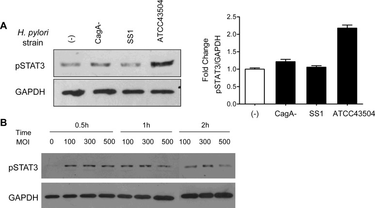
Figure 1. STAT3 was activated in H. pylori ATCC43504 infected AGS cells.
(A) AGS cells were treated with CagA+ H. pylori strain ATCC43504 and CagA-H. pylori strains respectively. Whole cell lysates were analysed by immunoblotting with anti-pSTAT3 (Tyr705) antibody. The pSTAT3 protein band intensities were quantified and normalized to GAPDH intensities (right panel). (B) The expression of pSTAT3 was induced in AGS after treatment with ATCC43504 at various MOI and co-culture time.

