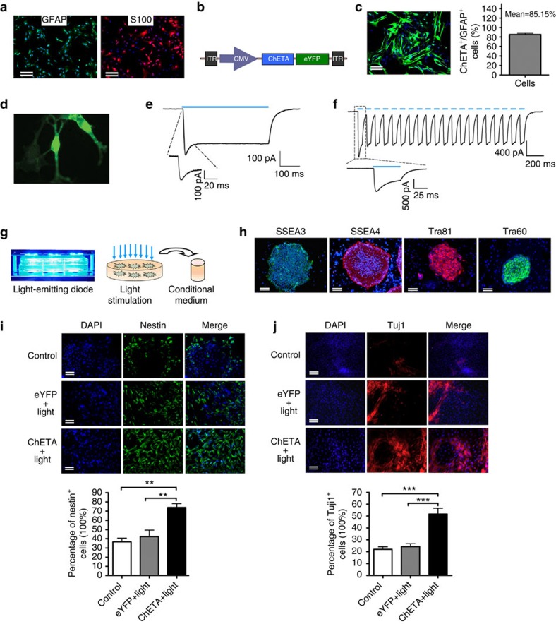
Figure 1. Light stimulation of astrocytes induced the neural differentiation of human embryonic stem cells.
(a) Immunofluorescence of glial fibrillary acidic protein (GFAP) and S100 on cultured rat-brain-derived astrocytes. Bar, 50 μm. (b) Schematic drawing of the CMV-ChETA-eYFP lentiviral vector construct. (c) Positive ChETA expression (green) was observed in the astrocytes transfected with CMV-ChETA-eYFP lentivirus (left panel). The percentage of ChETA-expressing astrocytes over the total GFAP-positive cells (right panel); mean±s.e.m. Bar, 25 μm. (d) Whole-cell patchclamp was used to record light-evoked photo currents from ChETA-expressing astrocytes. (e) Light stimulation (500 ms) induced depolarization and a recovery current. (f) ChETA channel current spike trains induced by light pulses (50 ms, 10 Hz). (g) The constructed blue-light- emitting diode was used to illuminate the cultured astrocytes and the conditioned medium was collected after the light stimulation. (h) Immunofluorescence of SSEA3, SSEA4, Tra81 and Tra60 on the human embryonic stem cells (hESCs); Bars, 50 μm. (i) Immunostaining of Nestin in Control, eYFP+light and ChETA+light groups. The percentage of Nestin-positive cells in the ChETA+light group increased compared with the Control and eYFP+light groups. Bars, 50 μm, (mean±s.e.m., n=10). (j) Immunostaining of Tuj1 in the Control, eYFP+light and ChETA+light groups. The percentage of Tuj1-positive cells in the ChETA+light group increased compared with the Control and eYFP+light groups. Bar, 50 μm, (mean±s.e.m., n=10). All analyses based on Student’s t-test, **P<0.01; ***P<0.001. The experiments were repeated three times with similar results.

