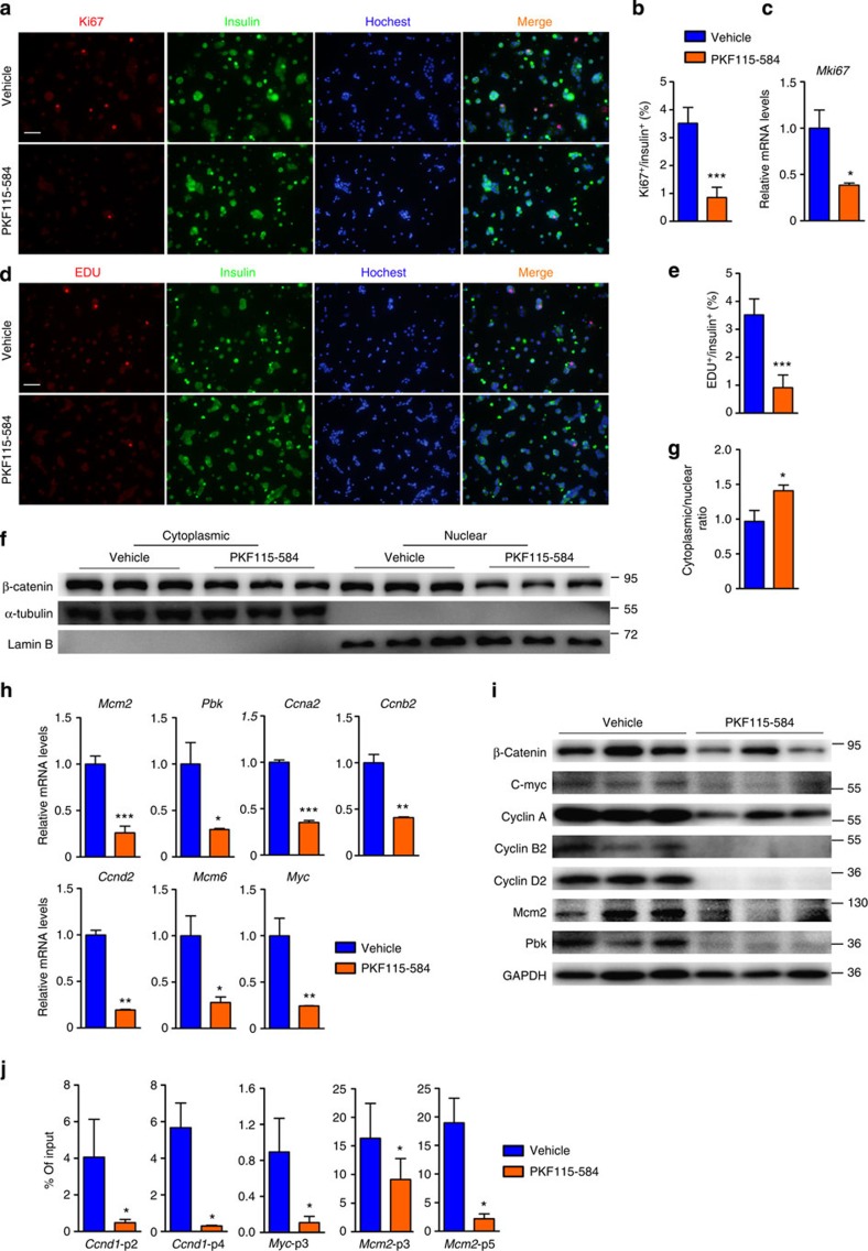
Figure 7. β-catenin antagonist inhibits replication of menin-null tumour cell and expression of proproliferative genes in vitro.
(a,b) Analysis of Ki67 staining on PKF115-584 (1 mM for 16 h) or vehicle-treated dispersed tumour cells from βMen1-KO mice (n=3). Scale bars, 100 μm. (c) Quantitative PCR (qPCR) analysis of Mki67 expression in PKF115-584- or vehicle-treated tumour cells from βMen1-KO mice (n=3). (d,e) Analysis of EdU staining on PKF115-584- or vehicle-treated dispersed tumour cells from βMen1-KO mice (n=3). Scale bars, 100 μm. (f,g) Western blot analyses of cytoplasmic and nuclear β-catenin in PKF115-584- or vehicle-treated tumour cells from βMen1-KO mice (n=3). (h,i) qPCR and Western blot analyses of Mcm2, Pbk, Ccna2, Ccnb2, Ccnd2, Mcm6 and Myc expression in PKF115-584- or vehicle-treated tumour cells from βMen1-KO mice (n=3). (j) ChIP assays were performed to show the binding of β-catenin at the promoters of Ccnd1, Myc and Mcm2 in vehicle or PKF115-584 (1 mM for 16 h) treated tumour cells from βMen1-KO mice (n=3). The pancreatic islets were isolated from 12-month-old βMen1-KO and βMen1/Bcat-KO mice. Sonicated cell lysates were incubated with β-catenin antibody for protein–DNA binding detection. Triplicate qPCR reactions for each sample showed the results from 660–400 bp (p2) and 2,750–2,552 bp (p4) of upstream regions of the Ccnd1 promoter; 1,030–885 bp (p3) of the Myc promoter; 712–585 bp (p3) and 1,428–1,326 bp (p5) of the Mcm2 promoter. All data are normalized against immunoglobulin-G control and expressed as percentage of input. The data represent the mean±s.d., *P<0.05, **P<0.01, ***P<0.001, Student’s t-test. The data shown represent three independent experiments.

