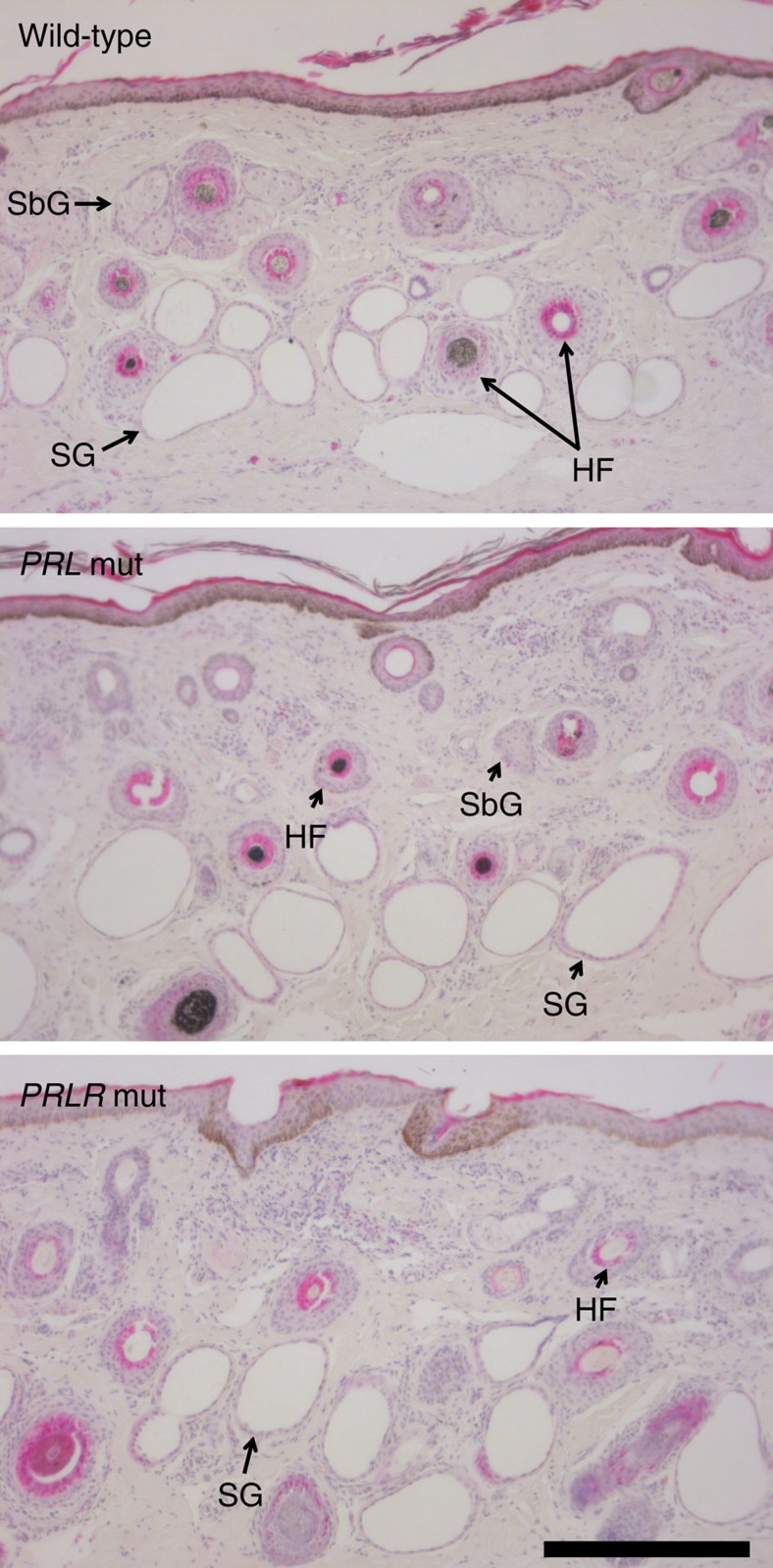
Figure 4. Skin histology of hairy and slick cattle.
Example haemotoxylin/eosin-stained skin sections at 100 × magnification representing wild-type (N=11), hairy (N=12) and slick (N=3) cows. The epidermis is top of field in each panel, sweat glands (SG), sebaceous glands (SbG) and hair follicles with and without fibre cross-sections (HF) are indicated. No qualitative or quantitative differences were observed between the different genotypes. Scale bar: 300 μm.

