
Figure 3. Properties of the excitation laser field.

(a) Intensity distribution and (b) polarization overlaps, 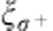 , ξπ and
 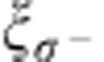 , of the excitation laser field in the vicinity of the nanofibre. The incoming field is modelled as a left-handed circularly polarized free-space plane wave that propagates in the +y-direction. The intensity in a is normalized to the incident intensity. The fibre radius is a=250 nm, the wavelength is λ=852 nm, and the local quantization axis is chosen along eϕ. The two alternative positions of the atoms are indicated by small crosses. From b, it is apparent that the excitation laser field is almost perfectly σ−- and σ+-polarized at the atomic positions, (r≈480 nm, ϕ=0) and (r≈480 nm, ϕ=π), respectively.