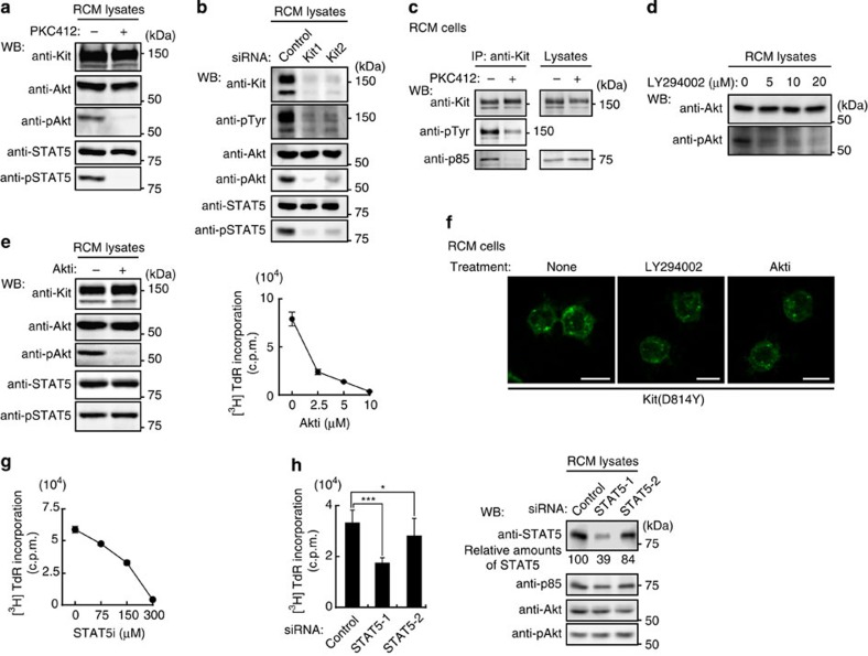
Figure 4. In mouse RCM cells, Akt and STAT5 must be permanently active for autonomous proliferation.
(a,b) Constitutive activation of Akt and STAT5 by Kit(D814Y). (a) RCM cells were treated with 1 μM PKC412 (Kit kinase inhibitor) for 24 h, lysed, then immunoblotted. (b) RCM cells transfected with control siRNA or Kit siRNAs were cultured for 20 h, then immunoblotted. (c) Association of Kit(D814Y) with PI3K. RCM cells were treated with 1 μM PKC412 for 24 h. Anti-Kit immunoprecipitates (left) and lysates (right) were then immunoblotted. NB: Kit(D814Y) was dissociated from p85 by PKC412 treatment. (d) Activation of Akt by PI3K. RCM cells were treated with the PI3K inhibitor LY294002. Lysates were then immunoblotted. (e) Role of Akt in proliferation of RCM cells. RCM cells treated with 10 μM Akti (Akt inhibitor) were cultured for 24 h. (Left) Lysates were then immunoblotted. (Right) [3H]-thymidine incorporation. Results (c.p.m.) represent means±s.d. (n=3). (f) Effect of LY294002 and Akti on Kit(D814Y) localization. RCM cells were treated with 20 μM LY294002 or 10 μM Akti for 24 h and stained with anti-Kit. Bars, 10 μm. (g,h) Role of STAT5 in proliferation of RCM cells. (g) [3H]-thymidine incorporation into RCM cells treated with the STAT5 inhibitor STAT5i for 24 h. Results (c.p.m.) are means±s.d. (n=3). (h) RCM cells transfected with control siRNA or STAT5 siRNAs (STAT5-1 and STAT5-2) were cultured for 24 h. (Left) Bar graph shows the levels of [3H]-thymidine incorporation into RCM cells. Results (c.p.m.) are means±s.d. (n=11). Data were subjected to one-way ANOVA with Dunnett’s multiple comparison post-hoc test. *P<0.05; ***P<0.001. (Right) Lysates were immunoblotted. Amounts of STAT5 are expressed relative to control cell lysate after normalization with p85.

