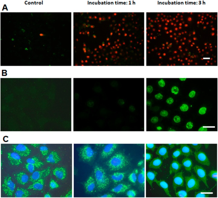
Figure 3.
Assays to characterize the type of cell death performed at different times after photodynamic treatments. (A) HeLa cells incubated with 1 µM ZnPc for 1 and 3 h, processed for the Annexin V/propidium iodide (ANV/PI) assays immediately after irradiation and observed in fluorescence microscopy under blue and green excitation light (overlay); (B) HeLa cells subjected to the same treatments, processed for TUNEL assay 3 h later and observed in fluorescence microscopy under blue excitation; (C) HeLa cells incubated with 1 µM ZnPc (1 and 3 h), processed for indirect immunofluorescence for cyt-c immediately after irradiation, counterstained with H-33258 and observed in fluorescence microscopy under UV and blue excitation light (merged image). Scale bar: 20 µm.

