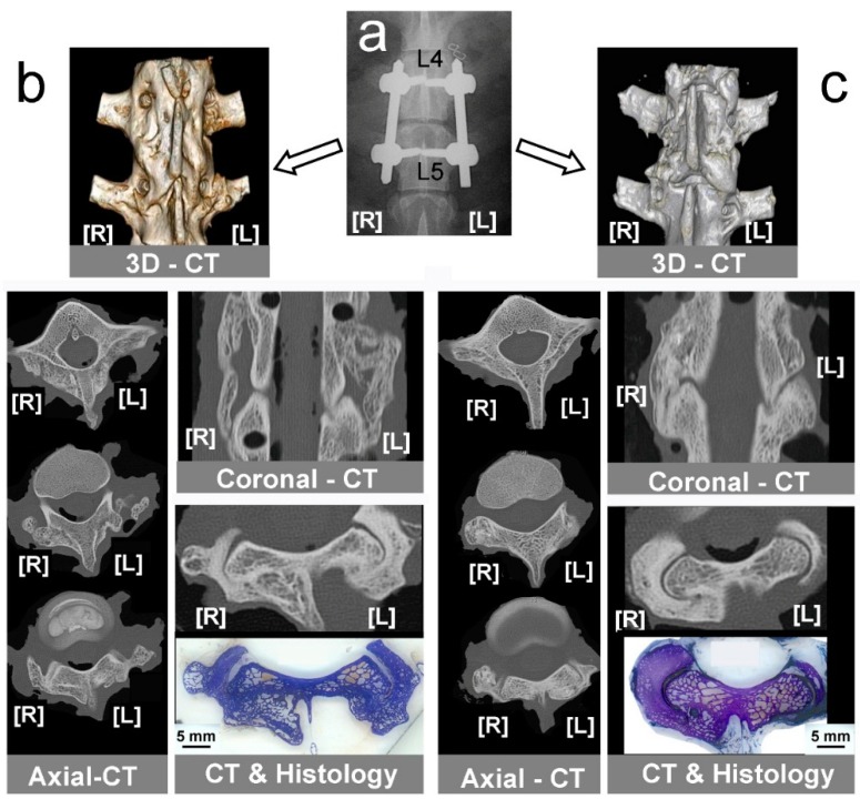
Figure 3.
Computed tomography (CT) diagnosis of instrumented lumbar fusion. (a) Control X-ray radiography taken after surgery, showing screws and rods used to fix L4–L5 vertebral bodies. [L], left side; [R], right side; (b) CT-study in bone graft groups showing 3D-CT reconstruction, axial-CT, a coronal-CT showing bone formation in both sides, and correlated axial-CT & histological section. [L] left side, Allo-group; [R] right side, Auto-group; (c) CT-study in mineral scaffold groups showing 3D-CT reconstruction, some axial-CT, a coronal-CT, and correlated axial-CT and histological section, assessing fusion only in right side. [L] left side, HA group; [R] right side, HA + MSCs group.

