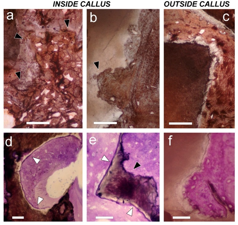
Figure 6.
Histology of mineral scaffold surfaces with deposited bone tissue. (a,b) On the left side (the HA group (a)), and on the right side (HA + MSCs group (b)), mineral scaffolds found inside the callus have an eroded contour (black arrows) that indicates previous osteoclastic resorption; (c) On the contrary, the mineral scaffold outside the callus on the right side (HA + MSCs group) does not; (a–c) von Kossa staining. Bar = 50 µm; (d,e) Inside callus, on left side; In the HA group (d), as well as on the right side (HA + MSCs group (e)), mineral scaffolds show a peripheral cement line (white arrows) that has been deposited before bone formation; (f) On the contrary, a cement line is not observed on the mineral scaffold surface when mineral scaffolds are found outside the callus on the right side (HA + MSCs group); in this case, bone tissue was formed by adding cells directly onto the mineral scaffold surface. Toluidine blue/pyronin G staining. Bar = 50 µm.

