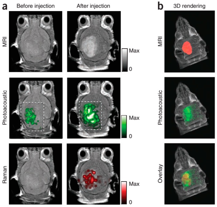
Figure 6.
Triple-modality detection of brain tumors in living mice with MPRs. (a) Two-dimensional axial MRI, photoacoustic and Raman images. The post-injection images of all three modalities showed clear tumor visualization (dashed boxes outline the imaged area); (b) A three dimensional (3D) rendering of magnetic resonance images with the tumor segmented (red; top), an overlay of the three-dimensional photoacoustic images (green) over the MRI (middle) and an overlay of MRI, the segmented tumor and the photoacoustic images (bottom) showing good colocalization of the photoacoustic signal with the tumor. (Reprinted from reference [52]. Copyright with permission from © 2012, Rights Managed by Nature Publishing Group).

