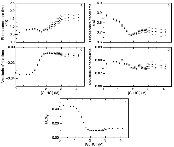
Figure 2.
Denaturant-dependencies of rise and decay times of acceptor fluorescence upon donor excitation track folding of apoflavodoxin labeled with A488 (donor) and A568 (acceptor). In all panels, black dots represent fitted values and grey triangles represent confidence limits. (a) The rise time of acceptor fluorescence and (b) decay time of acceptor fluorescence reveal the biphasic dependencies on GuHCl; (c) The amplitude of the acceptor fluorescence rise time (A−) changes in a monophasic manner as a function of GuHCl; (d) The amplitude of acceptor fluorescence decay (A+) is virtually constant as a function of denaturant concentration; and (e) The absolute ratio of the amplitudes of fluorescence rise and decay time (|A−/A+|) shows a monophasic dependence on denaturant concentration.

