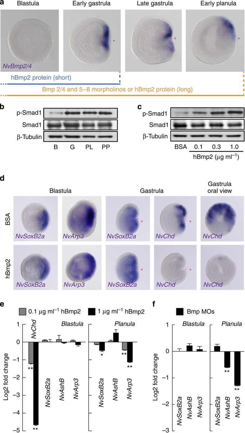
Figure 6. Bmp-independent early neurogenesis.
(a) WISH analysis of orally expressed NvBmp2/4 and scheme of hBmp2 treatment and of knockdown using morpholinos against Bmp2/4 and Bmp5–8. (b,c) NvSmad1 phosphorylation at the blastula (B), gastrula (G), planula (P) and primary polyp (PP) stages (b) and in blastula treated with increasing concentrations of exogenous hBmp2 protein (c). Smad1 and β-tubulin proteins were used as internal control. (d) WISH analysis of the effect of hBmp2 treatment (1 μg ml−1) on the expression of NvSoxB2a, NvArp3 and NvChordin (NvChd). (e) qPCR analysis of the early neurogenic genes at the blastula (left) and planula (right) treated with increasing concentrations of hBmp2. (f) qPCR analysis of early neurogenic genes at the blastulae and planula larvae injected with MOs against NvBmp2/4 and NvBmp5–8. Red asterisks denote the blastopore and future oral pole. Bars represent the mean±s.e.m. of three experiments. Black asterisks denote statistical significance using the Student’s t-test (*P<0.05; **P<0.01).

