Fig. 3.
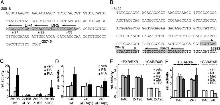
Drug induction of the two ADRES elements is mediated by NR REs. (A) Sequence of the 198-bp core ADRES element contained in hA6. The two contiguous potential NR REs (DR4 and DR5) are shown in gray. Arrows indicate the position of each of the three half-sites HS1, HS2, and HS3. Sequence numbers are relative to the ALAS1 transcriptional start site. (B) Sequence of the 240-bp core ADRES element of hA8. Numbering is relative to the ALAS1 transcriptional start site. Gray boxes show the predicted NR DR4 (1) and DR4 (2) REs. Arrows indicate the individual half-sites. (C) Reporter gene assays in LMH cells with two copies of wild-type and mutant (ΔHS1 to ΔHS3) 198-bp ADRES fragments. Cells were treated with vehicle, 400 μM PB, or 250 μM PIA. (D) LMH cell reporter gene assays with wild-type and mutant [DR4 (1) or DR4 (2)] 240-bp ADRES fragments. Treatment was analogous to treatment described for C.(E) Transactivation assays with hA6 fragment or two copies of the 198-bp ADRES. Reporter constructs were cotransfected into HepG2 cells with expression plasmids for human RXR-α and either human PXR or human CAR. To assess PXR-mediated activation, cells were treated with vehicle (0.1% DMSO), 10 μM Rif, or 400 μM metyrapone (Met). For human CAR, cells were treated with vehicle (0.1% DMSO) or 10 μM EE2. (F) Transactivation assays with hA8 fragment or the 240-bp ADRES contained within the hA8 fragment. Cells were treated as described for E. Data represent the mean of three independent experiments plus one SD.

