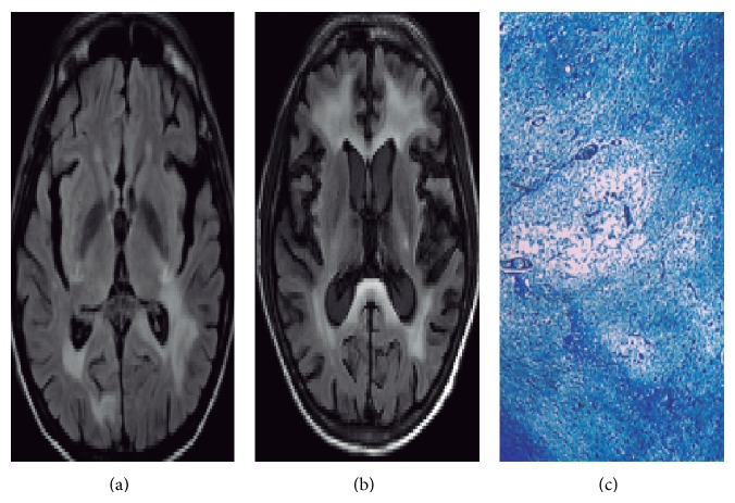
Figure 1.
Sequential MRI of the brain and myelin stain. Axial FLAIR images show hyperintensity in patches of subcortical white matter localized in parietal and occipital lobes (a). Premortem axial FLAIR images show increased lesions in bilateral frontal lobes without mass effect after treatment with maraviroc (b). Kluver-Barrera stain (10x) for myelin revealed brainstem zones of demyelination and enlarged oligodendrocytes with hyperchromatic nuclei and bizarre astrocytes (c).

