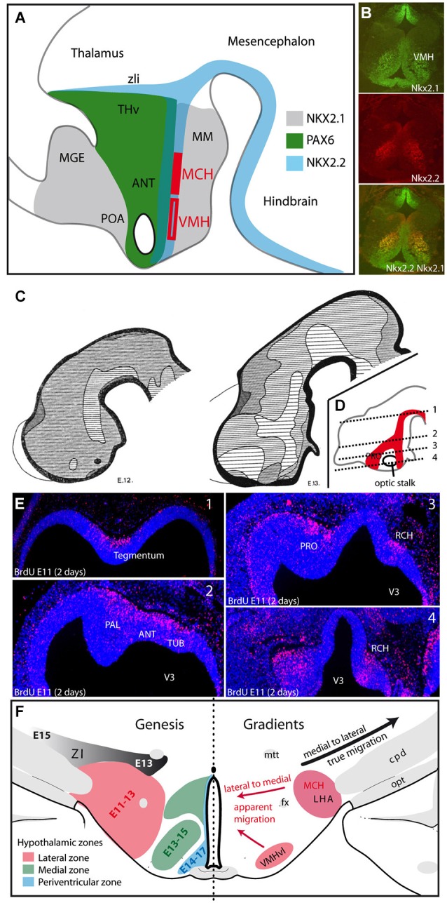
Figure 2.

(A) The distribution patterns of transcription factors agree well with the alar-basal divisions of the hypothalamus into preoptic, anterior and posterior/postoptic regions. However, these patterns extend outside the borders of the hypothalamus, involving the ventral telencephalon, prethalamus-ventral thalamus and ventral midbrain. MCH neurons and neurons of the VMH are generated from Nkx2.1/Nkx2.2 expressing neuroepithelial zones in the postoptic region. (B) Based on immunohistochemical analysis of a horizontal section of an E15 rat embryonic hypothalamus, the VMH clearly express both Nkx2.1 and Nkx2.2. (C) Figures from Keyser (1972) illustrating the differentiation of the early neurogenic zone in E12 and E13 Chinese hamster embryos: the first appearance of a longitudinal zone in the ventral mesencephalon and dorsal hypothalamus correspond to the cell cord (A). At E13, neurogenesis involved larger regions in the preoptic/ventral telencephalon; in other figure from Keyser that is not shown here, this author observed these regions forming one single continuum (as in D). This continuum takes the shape of an inverted Y. (D,E) Figure adapted from Croizier et al., 2011 illustrating the distribution of neurons generated at E11 on an E13 rat embryo. BrdU was injected into the pregnant dam at E11, and embryos were taken 2 days later at E13. BrdU was detected by immunohistochemistry on horizontal sections. The distribution pattern of these nuclei is schematized on a sagittal section in (D). BrdU-labeled nuclei follow an inverted Y pattern. In (E) pictures are arranged from dorsal (1) to ventral (4). (F) Gradients of neurogenesis in the ventral diencephalon. Left side: Schematic representation of the neurogenic gradients in the hypothalamus and prethalamus-ventral thalamus, as described by Altman and Bayer (1986). The LHA is generated between E11and E13, the medial hypothalamus from E13 to E15 and the periventricular zone from E14 to E17. Note the medial to lateral gradient in the prethalamus-ventral thalamus (generated from E13 to E15). Right side: Drawing summarizing the gradients in the ventral diencephalon: lateral to medial gradients (red arrows) in the hypothalamus suggest the apparent or passive migrations of neurons in lateral territories (LHA or VMHvl), but the prethalamus-ventral thalamus requires the effective migration of cells away from the ventricular surface (black arrow). Abbreviations: ANT: anterior hypothalamic area; cpd: cerebral peduncle; fx: fornix; LHA: lateral hypothalamic area; MCH: melano-concentrating hormone expressing neurons; MGE: medial ganglionic eminence; MM: mammillary body; mtt: mammillothalamic tract; opt: optic tract; PAL: pallidum; POA: preoptic area; PRO: presumptive preoptic area; RCH: retrochiasmatic region; THv: prethalamus or ventral thalamus; TUB: presumptive tuberal hypothalamic region; VMH: ventromedial hypothalamic nucleus; VMHvl: ventrolateral part of the VMH; ZI: zona incerta; zli: zona limitans intrathalamica; V3: third ventricle.
