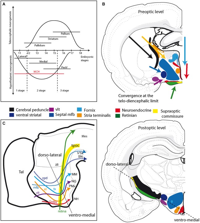
Figure 4.
(A) Schematic comparison of neurogenesis in the hypothalamus and telencephalon in the rat (see text for details). Neurogenesis in the hypothalamus is described as involving three stages: an early stage that produces only the lateral zone; a second that is concomitant to neurogenesis in the telencephalon and produces neurons in all hypothalamic longitudinal zones, but mostly the medial; a late third stage that concerns mainly periventricular zone neurons. Note that MCH neurons are produced during all three stages. (B) Illustration of the primary fiber tracts in the hypothalamus; these tracts originate in the telencephalon or retina and converge at the diencephalon-telencephalon limit at preoptic level (top drawing). In the postoptic hypothalamus (bottom drawing), these tracts are all aligned according to an axis determined by the dashed line. See text and supplementary information for details. (C) Schematic representation of the primary descending tracts in the ventral prosencephalon on a sagittal view of the embryonic brain. The tracts are topographically organized. Descending pathways from the telencephalon end in more posterior regions as they are distributed more dorsally in the hypothalamus. The dashed line recalls the ventro-medial/dorso-lateral axis, as in (B). Abbreviations: cpd: cerebral peduncle; fx: fornix; GnRH: gonadotropin-releasing hormone; MCH: melano-concentrating hormone; mes: mesencephalon; mfb: medial forebrain bundle; MM: mammillary body; NG: nucleus Gemini; NH: neurohypophysis; periV: periventricular; PMv: ventral premmamillary nucleus; SN: substantia nigra; st: stria terminalis; tel: telencephalon; tpoc: tractus postopticus; vlt: ventrolateral hypothalamic tract; VTA: ventral tegmental area; zli: zona limitans intrathalamica.

