Figure 3.
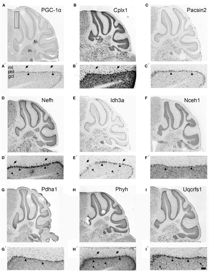
Neuroanatomical mRNA localization of PGC-1α and its dependent genes in the cerebellum. Sagittal in situ hybridization images of PGC-1α (A), Cplx1 (B), Pacsin2 (C), Nefh (D), Idh3a (E), Nceh1 (F), Pdha1 (G), Phyh (H), and Uqcrfs1 (I) transcript expression in the adult mouse cerebellum were collected from the Allen Brain Atlas (http://www.brain-map.org). All transcripts are highly expressed in the Purkinje cell layer (PCL; arrowheads) and the fastigial (FN) and interposed (IN) deep cerebellar nuclei. Most transcripts also exhibited expression in the molecular layer (ML; arrows), and Cplx1, Nefh, Phyh mRNA exhibited expression in the granule cell layer (GCL; concave arrowheads). Boxed portion in A represents area of higher magnification images in A′–I′. Scale bar = 525 μm for A–I and 100 μm for A′–F′.

