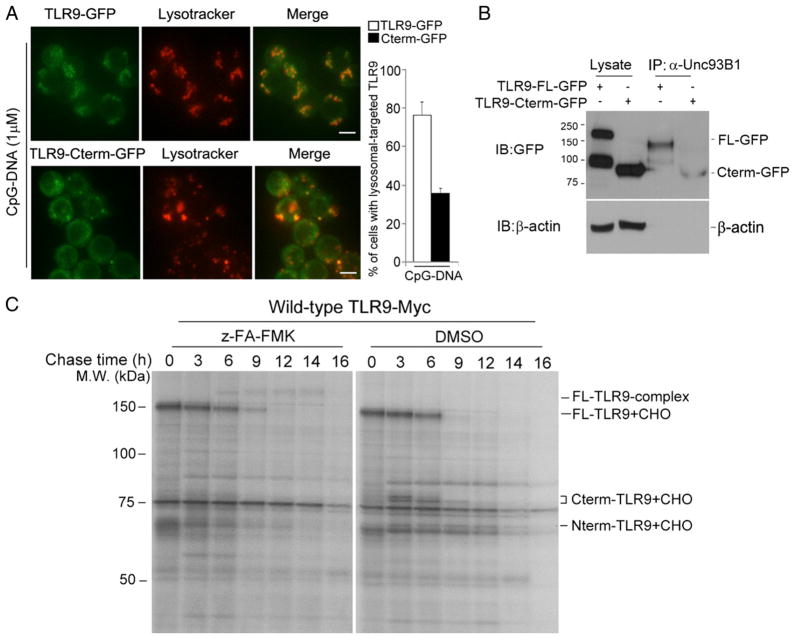
FIGURE 6.
The N-terminal region of TLR9 has distinct functions based on cellular localization. (A) RAW macrophages stably expressing either wild-type TLR9-GFP or C-terminal TLR9 fragments tagged with GFP (TLR9 Cterm-GFP) were stimulated with 1 μM CpG-DNA for 2 h, washed, and imaged after an additional 3 h with LysoTracker. Quantitative analysis of lysosomal-targeted TLR9 (right graph). Values represent the average percentage of cells with lysosomal-targeted TLR9 (n = 40–60 cells). Scale bars, 5 μm. (B) RAW macrophages expressing either wild-type TLR9-GFP or recombinant Cterm-GFP were lysed with 1% digitonin lysis buffer. Unc93B1 proteins were immunoprecipitated using the anti-Unc93B1 Ab. Immunoprecipitated samples were resolved by SDS-PAGE and analyzed using the anti-GFP Ab. (C) Wild-type TLR9-Myc–expressing macrophages were labeled with [35S]methionine/cysteine for 1 h and chased for the indicated times. Metabolically labeled cells were lysed with 1% digitonin lysis buffer, and lysates were immunoprecipitated using the anti-Myc Ab. Data are representative of three independent experiments.

