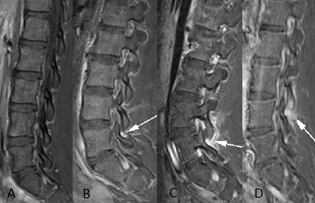
Figure 1.
Sagittal contrast-enhanced T1-weighted fat-saturated images of the lumbar spine in 4 different patients demonstrate the grades of apophyseal synovitis. A, Normal apophyseal joint. B, Abnormal enhancement (arrow) is confined within the apophyseal joint capsule indicating a grade 1 synovitis, clearly showing inflammation as compared to the normal apophyseal joint (A). C, Grade 2 synovitis with intraarticular synovial enhancement and periarticular high signal (arrow). D, Marked synovial enhancement and bone edema of the inferior articular process (arrow), indicating a grade 3 synovitis.

