Figure.

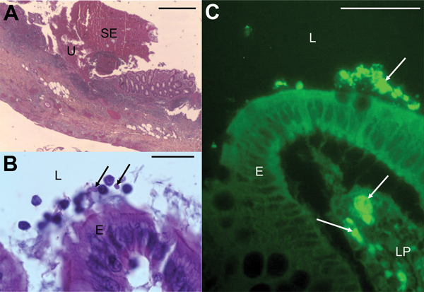
Micrographs showing histopathologic examination of appendix samples from a child who had peritonitis, Casablanca, Morocco, 2013. A) Ulceration (U) covered with suppurative and fibrinous exudates (SE) (hematoxylin-eosin stain). Scale bar indicates 200μm. B) Blastocystis parasites (arrows) in the lumen (L), and at the surface of the epithelium (E) (hematoxylin-eosin stain). Scale bar indicates 20μm. C) Blastocystis parasites (arrows) in the lumen, at the surface of the epithelium and in the lamina propria (LP) of the mucosa (immunofluorescence labeling with anti-Blastocystis ParaFlorB antibody). Scale bar indicates 50μm.
